
It is estimated that more than 30 million individuals in the India live with diabetes, and roughly 7.7 million of them have diabetic retinopathy, maki
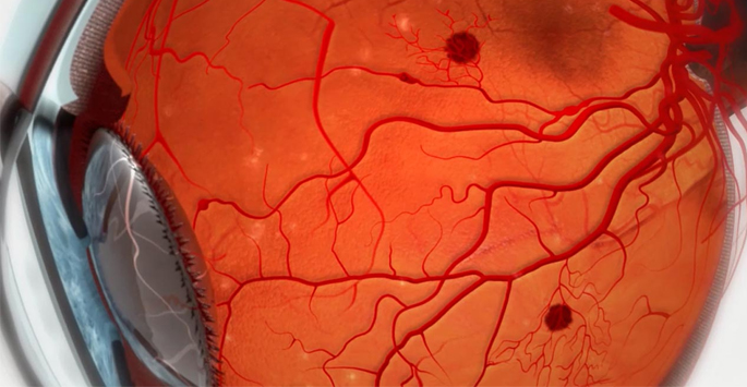
The retina is the light-detecting part situated in rear of the eye. It consists of nerve cells (neurons), specific cells called photo receptors that are associated with directly detecting light and blood vessels. The ability of retina to detect the light requires energy, which is subject to the oxygen provided by blood flowing through the vessels. In diabetes, increased glucose levels harm the vessels of the retina. These harmed vessels release fluid, leak blood and don’t provide sufficient oxygen to the retina, prompting retinal ischemia. Subsequently, retinal cells start to die and the retina doesn’t work as expected. Moreover, diabetes additionally harms the neuron of the retina directly. Together, these impacts cause diabetic retinopathy. Vision impairment related with diabetic retinopathy may at first influence focal vision because of a condition called diabetic macular edema. This fluid collection on the macula, a part of the retina responsible for sharp, focal vision, can prompt foggy vision and contortion of pictures. Progressed diabetic retinopathy is described by the arrangement of sporadic vessels that can bleed inside the eye, causing a sudden loss of vision. This results in an abrupt, drape like vision impairment as blood collects within the eye. Further deterioration of advance diabetic retinopathy can prompt retinal detachment, which requires critical careful mediation and can bring about long-lasting, irreversible vision impairment if not expeditiously treated.